Foot and ankle anatomy, conditions and treatments
When to see a doctor Summary The foot is an intricate part of the body, consisting of 26 bones, 33 joints, 107 ligaments, and 19 muscles. Scientists group the bones of the foot into the.

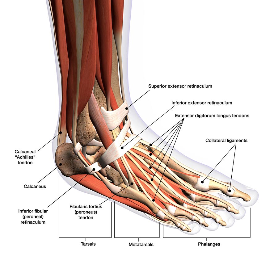
Foot and Ankle Musculoskeletal Key
The foot is the region of the body distal to the leg that is involved in weight bearing and locomotion. It consists of 28 bones, which can be divided functionally into three groups, referred to as the tarsus, metatarsus and phalanges. The foot is not only complicated in terms of the number and structure of bones, but also in terms of its joints.

Foot Description, Drawings, Bones, & Facts Britannica
Ankle anatomy The ankle joint, also known as the talocrural joint, allows dorsiflexion and plantar flexion of the foot. It is made up of three joints: upper ankle joint (tibiotarsal), talocalcaneonavicular, and subtalar joints. The last two together are called the lower ankle joint.
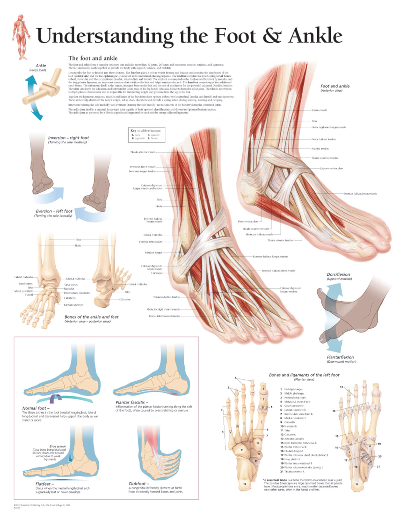
Understanding the Foot & Ankle Scientific Publishing
The 20-plus muscles in the foot help enable movement, while also giving the foot its shape. Like the fingers, the toes have flexor and extensor muscles that power their movement and play a large.

Medial Muscles And Bones Of The Foot Sole Labeled Human Anatomy Diagram Stock Photo Download
The phalanges create the toes. Each toe consists of three separate bones and two joints, except for the big toe, which has only two bones — distal and proximal phalanges — and one joint, like the.
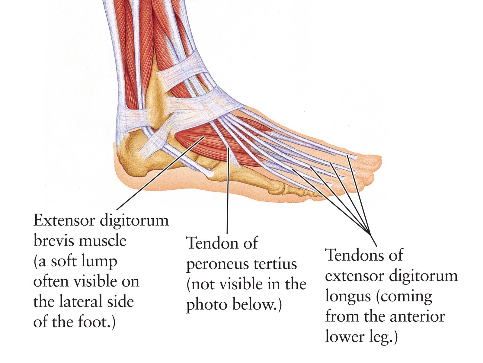
Human Anatomy for the Artist The Dorsal Foot How Do I Love Thee? Let Me Count Your Tendons
Common causes of foot pain include plantar fasciitis, bunions, flat feet, heel spurs, mallet toe, metatarsalgia, claw toe, and Morton's neuroma. If your feet hurt, there are effective ways to ease the pain. Some conditions specific to the foot can cause pain, less movement, or instability. Verywell / Alexandra Gordon.

Foot bones anatomical vector illustration labeled diagram Human body anatomy, Diagram, Vector
The bones of the foot provide mechanical support for the soft tissues; helping the foot withstand the weight of the body whilst standing and in motion. They can be divided into three groups: Tarsals - a set of seven irregularly shaped bones. They are situated proximally in the foot in the ankle area. Metatarsals - connect the phalanges to.

bones of the foot Bones of the Leg and the Foot skeleton of the hindlimb Documentation for
Foot Diagram with Labels The foot is situated at the distal part of the lower limb. It is one structure that has undergone several evolutionary changes. The foot of humans has changed from grasping to a supporting structure. It supports the whole body weight while standing and also plays a vital role in locomotion.
.jpg)
33 Label The Foot Labels 2021
Figure 1: Bones of the Foot and Ankle Regions of the Foot The foot is traditionally divided into three regions: the hindfoot, the midfoot, and the forefoot (Figure 2). Additionally, the lower leg often refers to the area between the knee and the ankle and this area is critical to the functioning of the foot.

Foot & Ankle Bones
The foot ( pl.: feet) is an anatomical structure found in many vertebrates. It is the terminal portion of a limb which bears weight and allows locomotion. In many animals with feet, the foot is a separate [clarification needed] organ at the terminal part of the leg made up of one or more segments or bones, generally including claws and/or nails.

anatomy of the foot Ballet News Straight from the stage bringing you ballet insights
Foot Anatomy and Biomechanics. base of the 5th metatarsal (lateral band), plantar plate and bases of the five proximal phalanges. plantar support is by the superficial and deep inferior calcaneocuboid ligaments. broad insertion over the lateral aspect of the lateral sesamoid and lateral aspect of the base of the proximal phalanx.

Ankle and Foot Pain Massage Therapy Connections
Foot Bones: Forefoot. The forefoot consists of 19 bones; 5 metatarsal bones and 14 phalanges. The big toe has 2 phalanges bones, while the remaining four have 3 phalanges each. The 1st metatarsal is the shortest and thickest of the metatarsals, and it is designed to take up to 40% of your body weight in standing, which rises to 70% when walking.

Structure of the human foot bone Royalty Free Vector Image
Use these bones of the foot quizzes to master your identification skills. Overview of the bones of the foot and their divisions into the hindfoot, midfoot and forefoot. With a total of 26 bones in each foot, learning the bony anatomy of the foot is no piece of cake. That is, the memorization aspect.

Foot and Ankle Musculoskeletal Key
It is made up of over 100 moving parts - bones, muscles, tendons, and ligaments designed to allow the foot to balance the body's weight on just two legs and support such diverse actions as running, jumping, climbing, and walking. Because they are so complicated, human feet can be especially prone to injury.
.jpg?response-content-disposition=attachment)
Foot Bone Diagram resource Imageshare
Anatomy of the foot Added to Saved items It may surprise you to know that the foot is one of the most complicated structures of the body. It contains a lot of moving parts - 26 bones, 33 joints and over 100 ligaments. Such complexity is necessary because the foot is required to do many different activities such as walking, running and climbing.

Chart of FOOT Dorsal view with parts name Vector image Stock Vector Image & Art Alamy
Foot Bones - Names, Anatomy, Structure, & Labeled Diagrams Foot Bones Humans have 26 bones in each foot that are classified into three groups - tarsals, metatarsals, and phalanges. These bones give structure to the foot and allow for all foot movements like flexing the toes and ankle, walking, and running.